Premium Only Content

Craniofacial Imaging in Orthodontics
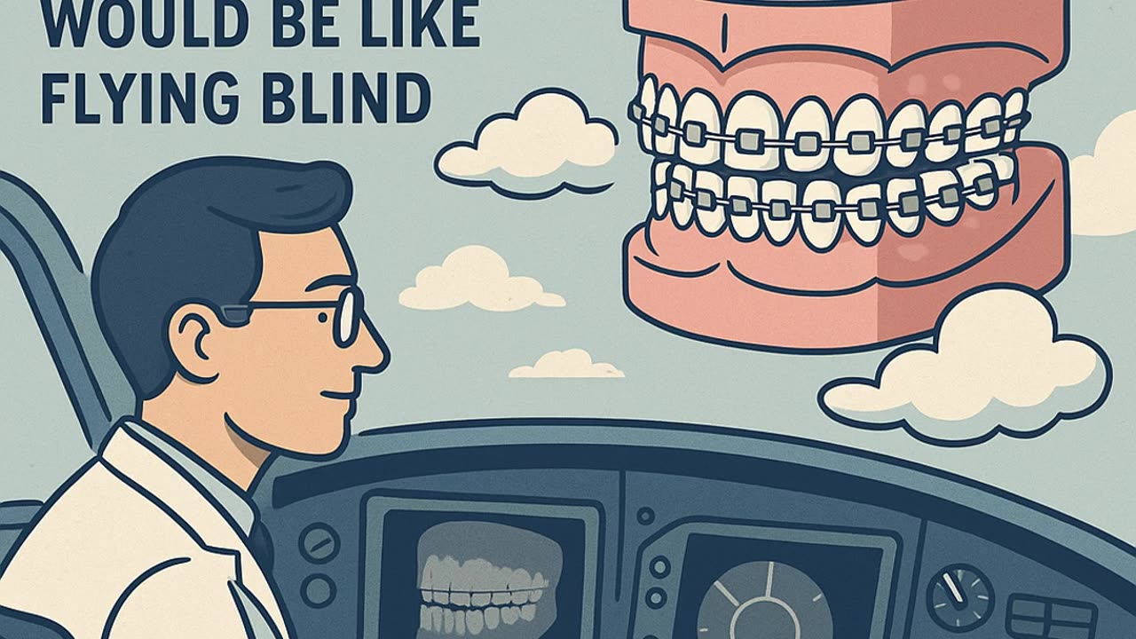
Imaging is the backbone of orthodontic diagnosis and treatment planning. But how do we move from classic 2D cephalometry to cutting-edge 3D imaging like cone-beam CT?
In this lecture, we break down the evolution and current practice of **craniofacial imaging in orthodontics**. You’ll learn:
* The history and limitations of traditional cephalometry
* Panoramic, periapical, and tomography applications
* How MRI helps in TMJ evaluation
* The rise of **cone-beam CT (CBCT)** and its advantages in orthodontics
* Digital imaging, stereophotogrammetry, and 3D patient-specific models
* How these tools change diagnosis, treatment planning, and outcomes
Whether you’re a dental student, orthodontic resident, or practitioner refreshing your knowledge, this video gives you a **clear, step-by-step guide** to modern orthodontic imaging.
📌 Stay tuned for future lectures in the orthodontics series.
👍 Like & Subscribe for more dental education content.
-
 2:17:46
The Connect: With Johnny Mitchell
4 days ago $17.00 earnedA Sitdown With The Real Walter White: How An Honest Citizen Became A Synthetic Drug Kingpin
96.3K2 -
 2:40:08
PandaSub2000
1 day agoDEATH BET | Solo Episode 01 (Edited Replay)
21.6K1 -
 9:41
9:41
Blabbering Collector
2 days agoHarry Potter Vintage Christmas Merch By Realtec Canada!
5.9K -
 LIVE
LIVE
Lofi Girl
2 years agoSynthwave Radio 🌌 - beats to chill/game to
841 watching -
 3:29:19
FreshandFit
14 hours agoMilo Yiannopoulos & Akademiks Find Out Who This Girl Smashed...
233K193 -
 2:08:35
Badlands Media
14 hours agoDevolution Power Hour Ep. 412 - Monroe Doctrine, Durham Rug, Income Taxes, and MORE!
87.1K15 -
 2:09:46
Inverted World Live
8 hours agoNASA Hints at Life Beyond Earth | Ep. 150
86.4K7 -
 19:01
19:01
Stephen Gardner
13 hours agoAlex Jones: ‘Trump Is About to Do Something MASSIVE!'
48.4K124 -
 5:14:39
Drew Hernandez
1 day agoCANDACE OWENS ACCEPTS TPUSA INVITATION TO DISCUSS SUSPICION BEHIND CHARLIE KIRK ASSASSINATION?!
50.6K21 -
 2:52:10
TimcastIRL
8 hours agoDrunk Raccoon Becomes Top US Story After Getting Plastered, Passing Out In Bathroom | Timcast IRL
237K123